 Skip to content


MRI scans are generally safe for people who have had scalp micropigmentation, and serious reactions are rare. The one thing to do without exception: tell the radiology team about your SMP before the scan, and speak up immediately if you feel unusual warmth or tingling once you’re in the machine. Guidance from Health Canada Safety Code 26 and a prospective study in the New England Journal of Medicine both support this verdict.
TL;DR:
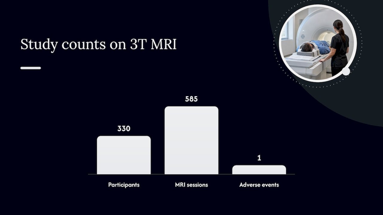
- The closest human evidence comes from a tattoo study, which recorded one mild reaction across 585 MRI sessions and estimated adverse event risk at 0.17% to 0.30%.
- Before scanning, list your SMP treatment date and location, share pigment details if available, and tell the technologist if the exact formula is unknown.
- Warmth or tingling can arise from skin contact, cables, coils, positioning, or pigment; report symptoms immediately so staff can pause or adjust the scan.
- Afterward, seek prompt follow up for blistering, persistent burning, spreading redness, or visible pigment changes; brief warmth that fades within minutes is typical.
Table of Contents
- What causes MRI interactions with SMP pigments
- What the research and safety guidance actually say
- Before, during, and after your MRI: what to do if you have SMP
- How common are reactions, and what do they feel like
- After the MRI: what’s normal and what needs follow-up
- How we document and protect every treatment we provide
- Does SMP cause lasting skin changes after an MRI?
- Do all SMP pigment colors carry the same MRI considerations?
- How should providers talk to clients about MRI compatibility?
- What if a doctor says MRI isn’t an option for you?
- A clinician’s note on MRI and SMP
- How we help you prepare for an MRI after SMP
- FAQ
- Sources
What causes MRI interactions with SMP pigments
The sensations some people report during MRI are not really about SMP as a whole. They trace back to specific ingredients and to the physics of the scanner itself.
Most pigment formulas used in scalp micropigmentation are designed to be inert, but older or lower-grade inks can contain trace metallic impurities, particularly iron oxides. These particles can respond to magnetic fields in ways that pure carbon-based pigments do not. An MRI machine itself works through three overlapping fields: a static magnetic field that aligns particles in tissue, a gradient field that shifts rapidly to build the image, and radiofrequency (RF) pulses that excite tissue and can generate localized heating or small induced currents in any conductive material.
Pigment is only one possible factor, and often not the main one:
- Skin-to-skin contact (arms touching sides, for example) can form a conductive loop and cause a burning sensation unrelated to any pigment.
- Monitoring cables or improperly placed coils are a more common cause of reported MRI burns than tattoo or SMP ink.
- Body position and padding inside the scanner affect how RF energy distributes across skin.
What the research and safety guidance actually say
The evidence on tattoos and MRI, which is the closest proxy available for SMP pigment, is reassuring but not absolute.
A prospective study published in the New England Journal of Medicine followed 330 participants through 585 MRI sessions and found only one mild tattoo-related reaction that led to a scan being stopped. The authors estimated the probability of a tattoo-related adverse event at roughly 0.17% to 0.30% under the study’s conditions on 3T scanners.
An animal study using ultra-high-field 9.4-T MRI found no burns and only minimal heating in inorganic tattoo specimens, a field strength well above the 1.5 to 3T machines used in most clinical settings (J-STAGE). Histologic changes were negligible. Because this was a mouse model at an unusually high field strength. It cannot be treated as direct proof of human outcomes, but it does suggest that even aggressive test conditions don’t reliably produce tissue damage.
Health Canada Safety Code 26 frames MRI exposure to static, gradient, and RF fields as a case-by-case risk-benefit decision rather than a blanket restriction, and it specifically instructs that any patient who feels discomfort near a medical device or foreign material should alert the operator so the scan can be stopped. A broader review of the literature on tattoos and MRI notes that:
- Reported sensations are usually transient and resolve without treatment.
- Quantitative data remain limited because study conditions vary between institutions (NCBI).
Before, during, and after your MRI: what to do if you have SMP
A few simple steps cut the uncertainty down to almost nothing.
- Declare your SMP on the intake form the same way you would a tattoo, and note the approximate date and location where it was done.
- Ask the technologist to flag your file so the team is aware before you enter the scanner room.
- If your SMP provider can supply pigment batch information or a safety data sheet, bring a copy or have it ready to email.
- Report any tingling, warmth, or tightness the moment you notice it. Staff can pause the sequence, adjust settings, or reposition you.
Technologists have several options available if a concern is flagged ahead of time, including lowering the specific absorption rate (SAR), changing the coil or pulse sequence, or simply watching the treated area more closely during the scan. Your job is simpler. Speak up the instant something feels off rather than waiting to see if it passes.
Pro Tip: Ask your SMP provider for a one-page summary with the treatment date and pigment details, and keep a copy with your health records so it’s ready whenever a scan gets scheduled.
How common are reactions, and what do they feel like
The NEJM data put the probability of any tattoo-related adverse event at roughly 0.17% to 0.30% under study conditions, a very small fraction of scans (NEJM). That figure comes from tattoo research rather than an SMP-specific trial, but it is the closest available benchmark given the similarity in pigment chemistry.
When something does happen, it tends to be minor:
- Brief tingling or a pins-and-needles feeling over the treated area.
- A sensation of mild warmth or tightness that fades within minutes of the scan ending.
- No lasting mark, blister, or skin change in the vast majority of reported cases.
Neither the human study data nor the animal research supports a real risk of serious burns, though every scan still deserves an individual look by the radiology team rather than an assumption either way.
After the MRI: what’s normal and what needs follow-up
Most people notice nothing at all once the scan is done, or at most a short-lived warmth that fades on its own. Watch instead for signs that fall outside that pattern: blistering, burning that persists past the appointment, spreading redness, or any visible change to the pigment itself. Our guide to SMP side effects covers what’s typical healing versus a warning sign in more detail.
If something feels wrong during the scan, tell the radiology staff right away since they can intervene immediately. For anything that shows up afterward, start with the radiology department for the acute event, then follow up with your SMP provider or physician. Keep the date of your original treatment handy for either conversation. An uneventful MRI does not call for any change to your long-term SMP care.
How we document and protect every treatment we provide
We select pigments and apply them conservatively, aiming to keep the long-term appearance natural and limit impurities linked to sensations during MRI, as outlined in detail by Scalp Micropigmentation in London | GetHair. We keep a record of the procedure date and pigment details for every client treated.
If you have an MRI scheduled and want that information in hand, you can request documentation or pigment safety data. Contact your SMP provider before the appointment to obtain what your radiology team needs to feel confident proceeding.
Does SMP cause lasting skin changes after an MRI?
The available evidence, both the human tattoo data and the animal study at 9.4T, points toward no lasting skin damage in the overwhelming majority of cases. The 9.4-T study found negligible histologic change and only a small, temporary temperature rise in pigmented tissue, which is a reassuring sign given that most clinical scanners run at far lower field strengths of 1.5 to 3T (J-STAGE).
Where concern does arise, it usually centers on pigment migration or color shift rather than tissue injury, and there is no clinical evidence tying MRI exposure to that kind of change. A transient warm or tingling sensation during the scan, if it occurs at all, typically resolves within minutes and leaves no visible trace. If you notice blistering, persistent burning, or a change in the appearance of your scalp pigment after a scan, that is worth a prompt follow-up with your SMP provider or a physician, since those findings fall outside what the research describes as a typical outcome. For most people, though, an MRI is simply a non-event for their SMP, with nothing to monitor once the appointment is over.
Do all SMP pigment colors carry the same MRI considerations?
Pigment composition varies more than color does. The factor that matters for MRI is whether a formula contains trace metallic compounds, commonly iron oxides, rather than whether the final shade reads as dark brown, soft gray, or a lighter tone matched to finer hair.
Carbon-based pigments, which make up most modern high-quality SMP ink, tend to be more inert under magnetic exposure than older iron-oxide-heavy formulas. The 9.4-T animal study tested both iron-oxide and carbon black specimens and found only minor temperature differences between them, with neither producing burns or lasting tissue change. That said, there is no public registry that lets you check a specific commercial pigment’s exact mineral content against an MRI safety database, so the practical answer is less about memorizing shade names and more about knowing what’s in your ink. Reputable providers can tell you whether their pigments are iron-oxide-based or carbon-based, and that detail belongs in any documentation you share with a radiology department before a scan. If your SMP was done years ago at a clinic that no longer operates, it’s reasonable to simply tell the technologist you don’t have the exact formula and let them make standard precautionary adjustments.
How should providers talk to clients about MRI compatibility?
SMP artists and dermatologists are in a good position to remove uncertainty before it ever becomes a problem, simply by treating pigmentation like any other cosmetic tattoo during intake and aftercare conversations.
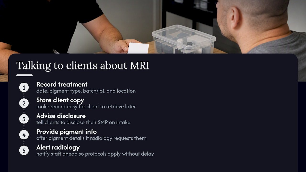
Best practice includes recording the treatment date, general pigment category (carbon-based versus iron-oxide-based, where known), and batch or lot information at the time of service, then making that record easy for the client to retrieve years later. A Manual on MR Safety used in radiology training emphasizes that documenting the date and location of cosmetic tattoos, and notifying staff ahead of a scan, are the steps that let a team apply any specialized protocol without delay. Providers should mention upfront, as part of standard aftercare guidance, that clients scheduling an MRI should disclose their SMP on intake forms and can request pigment details if a radiology department asks for them. This isn’t a legal requirement in most jurisdictions, but it closes the gap between what a client knows and what a radiology team needs, and it takes a provider only a few minutes per client to put in place.
What if a doctor says MRI isn’t an option for you?
MRI contraindications are rarely about SMP itself. They’re far more likely to involve other implants, devices, or patient-specific factors that a physician has already flagged, with SMP becoming a secondary question the radiology team asks about once those bigger issues are on the table.
When MRI is ruled out for an unrelated reason, imaging alternatives exist and your physician will choose based on what they need to see. CT scans use X-ray technology rather than magnetic fields and carry no pigment-related interaction at all. Ultrasound is another option for certain soft-tissue questions and involves no magnetic exposure whatsoever. Neither replaces MRI’s detail for every use case, so the choice depends entirely on what your doctor is trying to diagnose, not on your SMP history. If you’re ever told MRI is off the table, ask directly whether that decision relates to your pigmentation or to something else entirely. In nearly every documented case involving cosmetic tattoos or SMP, the answer is that the scan can proceed with standard precautions rather than being replaced altogether.
A clinician’s note on MRI and SMP
Scans go smoothly for the overwhelming majority of SMP clients we hear from, and the routine precautions work. Keep your treatment record, mention your SMP at intake, and say something the moment anything feels off. We’re glad to answer questions or provide documentation beforehand.
— James Christopher
How we help you prepare for an MRI after SMP
A record of pigment and treatment date is often kept for clients and can be provided when an upcoming MRI calls for it. That record can be useful to provide to a radiology department as it answers common intake form questions.

- Request a treatment summary or pigment details ahead of a scheduled scan.
- Review our scalp micropigmentation services to see our approach to pigment selection and documentation.
- Book a consultation through our treatment page if you’re considering SMP and want a provider who documents every session.
This article is general information, not a substitute for advice from a qualified doctor. Consult a qualified healthcare professional about your own circumstances before acting on anything here.
FAQ
Can I get an MRI scan after getting a tattoo?
Yes, MRI scans are routinely performed on people with tattoos, and a prospective NEJM study found adverse reactions to be rare under study conditions. Tell your radiology team about any tattoos or SMP beforehand so they can monitor you if needed.
What should I avoid before a brain MRI?
Follow your facility’s specific prep instructions, which typically cover metal objects, certain medications, and fasting requirements depending on the scan type. If you have SMP or a cosmetic tattoo, disclose it on your intake form regardless of what other prep steps apply.
Can my hair be wet for an MRI?
Wet or dry hair has no bearing on MRI safety, since the scan responds to magnetic properties in tissue and metal, not moisture. SMP pigment sits in the upper dermis and is unaffected by hair being washed or dried before an appointment.
What should I do after getting scalp micropigmentation?
Follow your provider’s aftercare instructions for cleaning and protecting the scalp during healing, and keep a record of your treatment date and pigment details. That documentation is useful well beyond healing, including for any future MRI appointment where a radiology team wants pigment information on file.
How common are reactions to MRI scans for people with SMP?
Research isn’t SMP-specific, but the closest available data, a prospective tattoo study, put the probability of an adverse reaction at roughly 0.17% to 0.30% under study conditions on 3T scanners (NEJM). When reactions occur, they’re typically mild tingling or warmth that resolves quickly.
Sources
- Guidelines on exposure to electromagnetic fields from magnetic resonance clinical systems – Safety Code 26
- Safety of Tattoos in Persons Undergoing MRI
- Multifaceted Evaluation of Ultra-high-field 9.4-T Magnetic Resonance Imaging after Inorganic Tattoos: An Animal Study
- NCBI review on MRI safety and tattoo interactions

